| Ogen In de pupil bevinden zich soms korrelvormige uitstulpingen van de rand van de iris. Het derde ooglid zit onder de binnenste ooghoek en kan zichtbaar gemaakt worden door zacht op de ooghoek te drukken. Bij een tetanusinfectie (klem) is het derde ooglid voortdurend geheel of gedeeltelijk zichtbaar. Door een tetanusinfektie kan een vergroeing van het regenboogvlies met de lens optreden. Een andere oogafwijking is maan(d)blindheid; een allergiese ontstekingsreaktie in het oog waardoor het paard niets meer kan zien. Door voeding krijgt het paard meestal de verontreinigende bacteriën binnen. Denk bijvoorbeeld aan uitwerpselen van ratten en muizen in het hooi en in de biks. In de binnenste ooghoek begint ook het traankanaal. Dit dient voor het vochtig houden van het oog. Het traankanaal loopt door de schedel heen naar het neusgat. Het kan verstopt raken, dan moet het doorgespoeld worden. In het hoofd bevinden zich diverse holtes: Oorspeekselklier: kan opzwellen en op droes lijken maar dat is het niet. Droes is een ontsteking van de lymfeklieren die begint in de keelholte. Door het paard aan de teugel te rijden kan de oorspeekselklier opzwellen en de luchtpijp dichtdrukken, waardoor het paard kan flauwvallen. De tong van het paard kan makkelijk scheuren. | |
    | |
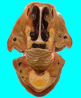 | tranverse section 1 This is a section through the canine region looking towards the diastema. The nasal vestibule, hard palate and incisive papilla are visible. Just visible on the left of the palate in the original specimen is the position of the canine tooth about to erupt, giving an approximate age of 3.5 years. This is difficult to see in the present image. Dorsal to the palate is the palatine venous plexus and the vomero-nasal organ. Ventro-laterally in the mandible are the roots of the lower canine teeth. |
| |
| tranverse section 2 This is through the diastema demonstrating the buccal vestibule laterally, the tongue centrally and the nasal conchae dorsally. Note the extreme development of the venous plexuses associated with both dorsal and ventral nasal conchae. | 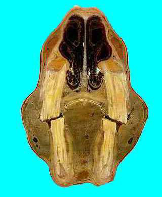 |
| |
| tranverse section 3 This is through the premolar teeth. In the upper jaw are PM3; in the lower are the forming roots of PM2. |
| |
| tranverse section 4 This is through the rostral maxillary sinus. The infraorbital canal is mounted on a septum that partly divides the cavity into arostral maxillary sinus laterally, and a ventralconchal sinus medially. Above thetongue is the caudal border of the hard palate. Dorso-medially is the nasal septum. | 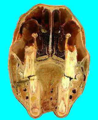 |
| |
 | tranverse section 5 This is through the caudal maxillary sinus. Demonstrating the paired frontal sinuses (medially) and the passage to the ventral conchal sinus lateral to these. Ventrally the facial vessel and parotid duct pass over the ventral border of the mandible. |
| |
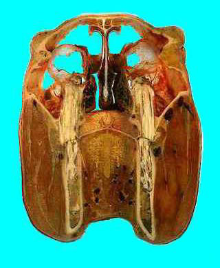
| tranverse section 6 This section passes through the caudal maxillary sinus and the orbits. The optic nerve is visible medial to the R. orbit. The nasopharynx, soft palate andoropharynx are visible along with (ventrally) some of the musculature connecting the hyoid region and the root of the tongue. The lingual process of the hyoid is in the ventral midline. Note the turbinates: these are the ethmoidals and in this regionthey are no longer covered by respiratory epithelium, unlike thosefurther forward. These have an olfactory epithelial lining. | 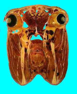 |
| |
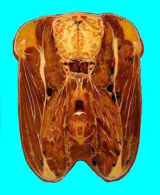 | tranverse section 7 This passes through the sphenoidal sinus and the thyrohyoid bone. Note that the epiglottis is visible above the soft palate: this differs from the situation seen in the longitudinal section where the epiglottis lies more caudally. Within the cranial vault rostral portions of the cerebral hemispheres maybe seen. |
| |
| tranverse section 8 The temperomandibularjoints are prominent. Ventrally in the larynx the vocalcords are visible. Note the pharyngeal entrance to the auditory tube (guttural pouch). This is a nice view showing the hippocampus forming the floor of the lateral ventricles, all within the cerebral hemispheres. | 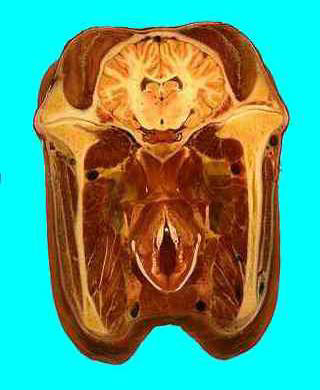 |
| |
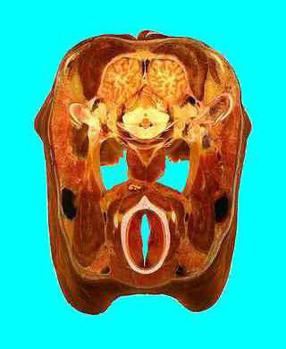 | tranverse section 9 This passes through the cartilages of the base of the ear. The paired occipital poles of the cerebral hemispheres and the vermis of the cerebellum lie above the large guttural pouches.The larynx is sectioned in the region of the vocal folds. The oesophagus is a muscular tube above the larynx to the left of the midline,and the large jugular veins are visible laterally. Note that cranial nerves 7 and 8 are closely associated with the middle-ear/petrous temporal region. |
| |
| tranverse section 10 This is a section through the occipital bone and, centrally, the medulla oblongata. The parotid gland lies laterally and superficial to the mandibular gland and jugular vein. The dark brown thyroid gland lies either side of the trachea. | 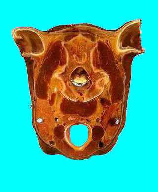 |
| |
School of Veterinary Studies Murdoch University Perth, Western Australia Horse Head Series
Background: This series of images was taken from 35 mm slides of original sections
of a deep-frozen horse head sliced with band saw and mounted in
Perspex pots in our Veterinary Anatomy Museum, within Murdoch
University.
The slides were scanned into Kodak Photo CD, imported into Adobe
Photoshop 2.5.1 for enhancement and cropping and finally saved as
JPEG files in thousands of colours with a maximum standard size of
480 x 640 pixels, using a Mac Quadra 840 AV.
The images were then resized to fit comfortably in the display
windows of WWW browsers such as Netscape and Mosaic. A format of
320x390 was used for the JPEG transverse sections, allowing a minimal
file size. The longitudinal section, which is a clickable map, is a GIF
image of size 600x390.
Credits:
The sections were originally prepared by Richard Krumins,
photographed by Geoff Griffiths, and annotated in June 1983 by
Associate Professor John Grandage, of Murdoch University's School of
Veterinary Studies.
The initial digitising, enhancement and posting was done with the aid
of a grant to Jim Cummins from the Australian Committee for the
Advancement of University Teaching, with the expert assistance of
Geoff Rehn from the Academic Services Unit of Murdoch University,
who developed the HTML as well as reworking the images for
publication on the Web.
Contacts:
Assoc. Prof. Jim Cummins cummins@possum.murdoch.edu.au
Geoff Rehn rehn@cleo.murdoch.edu.au