| coronar first aid heels P3 sequence sole diagnose | ||
| Laminated? Laminae are what bind the hoof wall to the foot bone. They are an example of tissue that is at once durable and vulnerable. They transfer from hoof wall to P3 (foot bone), and vice versa, all the force generated between horse and earth. At times this is a tremendous load. The laminae successfully carry out this task with apparent ease. |  | |
| The laminae of the horny wall are called epidermal or insensitive laminae. They are made of non-living keratinized cells. The laminae which interlock with the epidermal laminae are called dermal or sensitive laminae. The dermal laminae are living tissues with a perfuse vascular system. | ||
| The horse has primary and secondary laminae. The larger primary laminae have secondary laminae around their edges. I haven't counted the laminae, but those who have say there are approx. 600 primary laminae in the average horse's hoof. The surface area produced by this system is huge considering the space it occupies. |  | |
| The brown stuff is horn (insensitive) and the red stuff is flesh (sensitive). Lamellar circulation consists of a dense vascular network. Arteries branch out becoming smaller in diameter until at their smallest diameter they are called capillaries. They then become larger as they branch back into larger vessels now called veins. This process of artery becoming vein is called anastomosis. To anastomose is to communicate, in a liquid sort ofway. Arteries and veins can communicate at larger branches as well. This is by way of shunts or AVAs (arteriovenous anastomoses). The horse's foot has a circulatory autoregulation system. By opening and closing AVAs to periodically flush greater amounts of blood through the foot it can stay warm in extreme cold conditions. This autoregulatory system may be important both in preventing founder and in causing laminitis. Laminitis is not fully understood at this time. There are several theories about it's etiology. My field experience leaves me firmly convinced that inflammation is the most important factor in most cases. In those cases where inflammation was controlled, so was the pathology controlled. | ||
    | ||
| A Brief, Elementary Introduction to Laminitis or Founder 101 Lamina [L.], A thin flat layer or membrane. itis [Gr.], Suffix meaning inflammation of. Laminitis is a malady effecting the horse's foot. There are many causes of laminitis. Most of the laminitis we see is called Alimentary laminitis. Each different cause may be called by many other names. For example, one form of alimentary laminitis might be called grass founder , another called grain-binge. A mare who has not discharged all of the placenta after giving birth is at risk of getting laminitis, some call this retained placenta, while others might call this metritis. The important thing to know is that there are many reasons a horse might get laminitis and many different names people will use when they talk about it. The reason we are so concerned about laminitis is that it can leave permanent scars in the horse's feet.Laminitis will affect each horse differently (even each foot of the same horse), from the barely noticeable caseto the fatal case. It is important to think of laminitis as a symptom of other problems the horse is experiencing.Because we become aware of laminitis through the lameness it produces in the feet, if we only fix the feet weare ignoring the real problem, and the lameness may come back. Laminitis means inflammation of the laminae. The Laminae (pronounced Lamin-nee) are the tissues that connectthe hoof wall to the Coffin Bone. The Laminae are arranged in vertical rows beginning at the Hair line, or Coronary Band, continuing down to the ground. The largest percentage of Laminae are in the front or Anterior region of the hoof, where they are longest. They gradually become shorter as they approach the heels. The hoof has Sensitive and Insensitive Laminae. The Laminae connected to the hoof wall are the Insensitive Laminae. The Laminae connected to the Coffin Bone are the Sensitive Laminae. The Sensitive Laminae are vascular andinnervated. The Sensitive and Insensitive Laminae Interdigitate. Laminitis is often confused with Acute Laminitis or Founder. Founder, which is a maritime term meaning sink,means that the Sensitive Laminae holding the Coffin Bone in place are letting go. In mild cases of Laminitis we are not always aware of it's presence. As inflammation (Laminitis) increases, Edema accompanies it. At this pointwe may begin to see lameness. Because the Laminae are between a rock and a hard place (the hoof wall andthe Coffin Bone) they have nowhere to expand to accommodate the edema. So Lamellar edema displaces theblood in the Capillaries of the Sensitive Laminae causing Pressure Ischemia. If the Ischemia persists it causes the Laminae to die. This is called Acute Laminitis or Founder. The Laminae in the front of the hoof, which carry most of the weight of the horse, will stretch and tear allowingthe front part of the coffin bone to pull away from the hoof wall. This allows the Distal Border of the Coffin bone to drop to varying degrees. This is called "Rotation" (measured in degrees). In severe cases all the Laminae die, allowing the Coffin Bone to drop right through the bottom of the hoof. This is called Vertical Displacement or "Sinker". We can say a horse has "Foundered" when either Rotation or Vertical Displacement has occurred. | ||
| ||
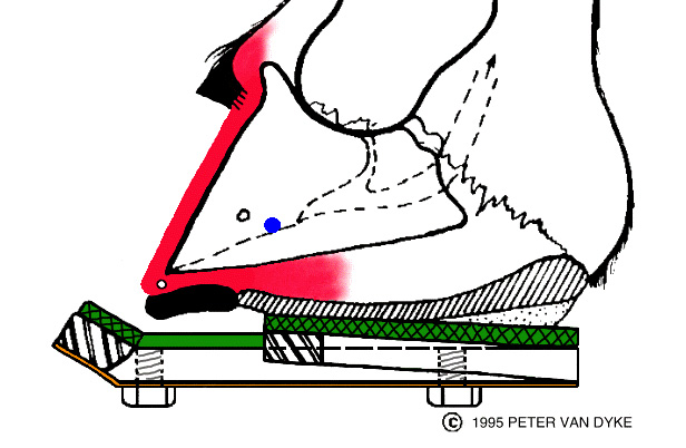
| The Coronary Band The pink stuff is the coronary corium and lamellar corium. The coronary corium is made of tissue thatproduces the cells that become the horn of the hoof wall. Injuries to the coronary band deep enough toinvolve the coronary corium can scar the corium causing it to produce low quality horn from the area ofthe scar. | 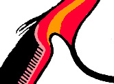 | |
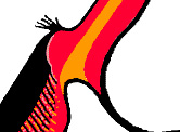 The correct position of P3 is vital to the coronary corium's ability to properly produce horn. The correct position of P3 is vital to the coronary corium's ability to properly produce horn. | ||
| ||
| First Aid For The Laminitic Horse This is a partial list of stages a Laminitic horse might experience. Each horse expresses the symptoms of Laminitis in his own way. A horse need not go through the progression I showhere. A horse might be minutes away from complete lamellar failure and just seem "not right". Another horse might beshow distress while not being in imminent danger of lamellar failure. The first horse is stoic and the latter a weenie.This is a disease that can be beat. If you do some research and act quickly you can get your horse through this easierthan you might think. Read everything you can get a hold of, talk to everyone you can, but Please be Proactive. Don't sitaround and wait to see what happens. If you don't think you are getting good advice, keep looking until you do. The Obel grading method is sometimes used to categorize laminitic horses by pain levels. More on Obel Grading First Stage Second Stage Third Stage Fourth Stage Frog Support The material I recommend for this kind of first aid is indoor-outdoor carpeting. Cut it in triangular pieces the shape and size of the frog (with a little extra at the base to go up over the heels), stack enough pieces on the frog that the pile projects a quarter to three eights of an inch beyond the bearing surface of the foot, and tape the support up around the foot.This gives you a compressible pad that supports but has a little give to it. If you don't happen to have indoor-outdoor carpeting, you can tape a roll of gauze under the frog instead; just don't use anything hard or unyielding, such as plywood, which could create additional problems by applying too much pressure. | ||
| ||
| Who said heels? Who cares about the heels if the toes aren't taken care of first? In many ways the toe defines the heel, so I don't think the heel should even be considered until the toe hasreceived all the work it might need first. Don't worry about the heels, do something about them. Shoe the bone. If P3 needs to be lifted behind, then lift it. Low or weak heels aren't good at supporting the weight of the horse they're under. They need to be replaced by heels that cansupport the horse. If P3 needs to be let down behind, then let it down. Shoe P3. The horn of the hoof wall tells a story. Listen to it. The hoof wall is not the story, but the teller. Get rid of what hinders the hoof, and utilize what helps the hoof. If you do this you are practicing farriery.
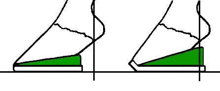
I didn't get these graphics quite the way I wanted them, but they'll do. The foot on the left is an example of the common use of wedge pads. This use of wedge pads is the reason they have received such bad press. This configuration will cause the heels to experience more stress than if just left alone. The foot on the right is more like the foot I want to see. Even better than wedge pads is heel reconstruction. If the money is there, heel reconstruction can do incredible things for a foot. Heel reconstruction is like crowning a tooth. A saturated fabric technique, much like fiberglass, is used to build on the existing heel. This process allows us to create the type of hoof we think will support the horse best. Reconstruction takes an otherwise deficient hoof and exerts forces on that hoof that mimic forces the hoof might have experienced had it had a more desirable shape originally. This can cause the new growth to follow the influence of the reconstruction,producing a more supportive natural hoof (with luck). Even when this doesn't happen, the reconstructed hoof can make for a sounder horse.
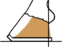
These drawings are all from the same base drawing. I used this drawing because it shows an ugly foot, one that can cause serious problems. I've over done the therapies a bit, but sometimes I will do it as shown when I feel I need to force the point. | ||
| ||
| The Coffin Bone P3 has a whole lot of stuff happening to it.Tendons, ligaments, laminae, cartilage, all kinds of good stuff attaches to it. |  | |
| The shape of the hoof is sometimes referred to as a split cone. The geometry of the split cone is important to how the horse's hoof functions. If you trace the outside of the hoof wall and the front part of P3 you will end up with shapes similar to these.
The lamellar surface area of P3 is about one quarter the surface area of the exterior of the hoof wall. P3 issmaller than most people imagine. Don't try this at home kids. To trace the front of P3 you have to take it out of the hoof (yuk!). But do try playingaround with the split cone. If you make a cone like the one above you can experiment with how the cone baresweight at various angles. You can see how certain angles seem better able to support weight than others. Youcan also see how too little angle doesn't absorb much shock. Watch how the "heels" move differently atdifferent angles. Sometimes they move outward away from each other, sometimes they move inward andforward. If you play with this long enough you should develop a reasonably good sense for how hoofconformation and trimming effects hoof function (form follows function or function follows form?). | ||
| ||
Sequence of Events During Laminitis
In the normal healthy hoof the sensitive and insensitive laminae provide a tight bond between PIII and the hoof wall. The laminae transfer most of the horse's weight from the skeleton to the hoof wall upon which he stands. At rest gravity is the main force acting on the lamellar bond. In motion, the pulling force of the Flexor Tendons is added to gravitational force. Trimming the hoof's toe as short as soundness allows and using a Rocker Toe Shoe reduces the length of the lever arm against which the Flexor Tendons pull. Depending on individual conformation, this can reduce hoof stress substantially. Any reduction in hoof stress is valuable to the sound or laminitic horse. | ||
| ||
During laminitis the laminae become inflamed. Inflammation is accompanied by edema (the attraction of intercellular fluids). There is a fixed fluid volume within the lamellar space. As edema collects, it displaces some other fluid; blood. If this process continues blood is displaced until insufficient blood flow exists to maintain life in the sensitive (live) laminae. The pressure caused by edema is also felt by the lamellar nerves, producing pain (this is not yet the pain from lamellar tearing). The pain level is indicativeof the edemic pressure level. It should be noted that the "Stoic" horse will show less pain than will the "Weenie" horse. The laminitic horse becomes the foundering horse when insufficient of blood flow allows lamellar tissue to die,causing the lamellar bond to fail. | ||
| ||
These early events can be called the Developmental stages of founder. Prior to the death of sensitive lamellar tissue, the lamellar bond isweakened. At this stage the laminae can stretch just enough to allow PIII to descend slightly, crushing capillaries in the solar corium below.Blood leaking from the crushed capillaries produces stains seen in the sole horn called "symmetrical subsolar hematomas". Similar stains mayalso be seen in the White Line. Because lamellar stretching can occur prior to the lamellar tearing associated with founder, red white lineand/or symmetrical subsolar hematomas can be present while the horse is still in the laminitic stage. In the case of the "Stoic" horse, these hematomas can appear with very little evidence of pain. Red white lines and symmetrical subsolar hematomas are important symptoms of laminitis and should not be ignored or confused with "Stone Bruises". The reverse is not true: A horse may be fight on the verge of foundering and show no signs of these stains. | ||
| ||
Once the laminae begin to die, the potential exists for rapid deterioration. At this point PIII will either rotate, sink, or both. This is "Founder" or "Acute laminitis". As the laminae are pulled apart the space created by the tearing may be filled with blood, serum, pus, or general gick. This is called the "lamellar wedge" This wedge must be removed before PIII can bereplaced to it's proper location. Now the horse has lost his opportunity for a scar free recovery without human intervention. | ||
| ||
Heart-Bar Shoe | ||
| ||
I fit a rim shoe to the foot just as I would normally do. With the shoe held against the foot I mark the shoe as per the crosshairs in the figure. I use these marks to locate the tip of the pressure bar for welding, adjust the height of the bar, and apply the shoe, and chanjo presto, thehorse is fixed (in your dreams). I am working on these drawings with some reluctance. The story these drawings tell is a very different story from the one this page is about. I'm using this short trip through founder/resection to highlight the importance of avoiding it. The story in these drawings is about advanced disease, or chronic disease. The day will probably not come that no horse will experience founder (for those that do this is important work), but each day more people are becoming familiar with the early signs of laminitis, reacting appropriately, and fewer horses are foundering. | ||
| ||
When PIII is allowed to rotate the laminae stretch, die, and become hard. This is the lamellar wedge. In order to restore health to the hoof capsule PIII must be replaced to it's pre-rotation position. The wedge occupies the space where PIII should be. So, the wedge has got to go. | ||
| ||
After the lamellar wedge has been removed PIII can be restored to It's pre-founder position.If the tissues that produce new growth are still healthy, horn and laminae should begin to grow normally or near normally (this is a big topic and here is not the place to get into it). | ||
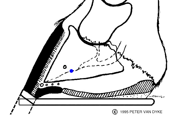
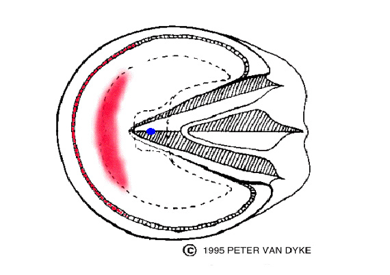
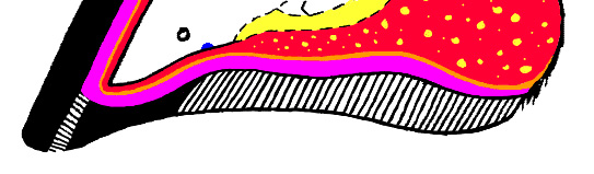
| Who says Horses don't have Soles The black represents horn. The purple represents papillae (solar and lamellar). The orange representscorium(solar and lamellar). The red represents connective tissue. The red with yellow represents the digitalcushion. The yellow represents the deep flexor tendon.
| ||
| ||
| A.A.E.P. BURNEY CHAPMAN AND GEORGE PLATT Thirtieth Annual Convention Dallas, Texas December, 1984 © Copyright 1984, Burney Chapman, George Platt L A M I N I T I S Although the foot is one of the most important parts of the horse's anatomy it is almost universallymisunderstood. Laminitis is generally agreed to be due to ischemia of the laminae, causing detachment of thethird phalanx from the horny wall. Founder is a maritime term meaning "sinking", founder is sinking of the thirdphalanx in the hoof, secondary to laminitis. Chronic laminitis is an "old founder" that has survived by somemeans. The third phalanx is demineralized with lytic areas being evident radiographically is usually badlydeformed and shows signs of chronic inflammatory changes. ANATOMICAL CONSIDERATIONS The union of the horny wall and the corium of the third phalanx is formed by the laminae. In the normal foot, the bond between these laminaecannot be broken. It is only after death of these structures that they can be separated. The sole is the horny covering or plate of horn on the bottom of the hoof. It is not usually as tough as the wall. Its origin is the solar matrix andit receives its blood supply from arteries branching from the terminal arch, drained by the subsolar venous plexus. The bond between thematrix and the sole is papillar, rather than laminar. They, too, cannot be separated in a healthy foot. Only when the foot is injured or in the caseof founder, can they be torn apart. The coronary band (coronet) produces the central laminar portion of the horny wall. The coronet is similar to the human cuticle and houses amass of minute capillaries. The horny wall starts below the coronary band and extends downward until it reaches the sole. The wall includingthese laminae is extremely important, as it is responsible for bearing most of the weight of the horse. The deep digital artery is the main blood supply to the third phalanx. It enters the hoof both medially and laterally at the bulb of the heel,passes through the foramen on either side of the semilunar crest, and forms the terminal arch. This artery is not protected by anything but thesole until it passes through the plantar foramen of the third phalanx where it anastomoses with its partner of the opposite side. If this arteryor its branches which perforate the wall of the third phalanx are destroyed, the horse will also be ruined. This is why a hoof cast, improperlyapplied, is so dangerous. Cases are known in which the blood supply was shut off and the digital artery was destroyed. The disposition of the vascular system of the complete hoof consists of two-thirds veins and one-third arteries. Two major vessels are thecircumflex vein and circumflex artery which run along and the distal peripheral edge of the third phalanx. At no point can sharp divisions berecognized. Each part unites and becomes continuous with the other. | ||
| ||
ETIOLOGY These conditions must be corrected before a favorable response can be expected. The major point to consider is that not only the cause must be determined, but also the extent of the damage to the feet. Measures need to betaken to correct this damage and steps must be taken to prevent further damage. The laminae swell in the early stages. Pressure increases between the hoof wall and the third phalanx. It is this pressure that causes theeventual necrosis, and it is this pressure that causes the rotation of the third phalanx. It is our opinion that the pressure from the deep flexortendon is a negligible factor in rotation of the third phalanx, unless there is a pre-existing flexor deformity, or unless the heels are cutabnormally low or the toe is elevated to create a secondary flexor deformity. The relationship between founder and flexor deformity is very complex and will be discussed later. Pre-existing conditions seen in many laminitic horses include: overfed, overweight and under-exercised; stall confinement; being shod tooyoung (as yearlings); having feet that are too small and flexor deformities. | ||
| ||
DIAGNOSIS If rotation of the third phalanx is not present, then one can supplement the diagnosis by running the finger down the middle of the leg to thecoronary band. If it encounters a depression behind the coronary band, straight vertical displacement of the bony column down through thehoof capsule (sinker) is likely. This is disastrous, as it does not show up radiographically except at the coronary band, and means that 100% ofthe laminae have separated. Furthermore, this straight vertical displacement is often missed. It will show only at the coronary band, anteriorlyon a lateral radiograph, and will have the appearance of the hoof capsule going proximally up the leg. Unless this sinking process is stopped, the hoof will slough. Due to the anatomy and weight of the equine species, when the bony column sinks,the coronary area of the hoof is jammed onto the coronary plexus, cutting off the blood supply to the plexus. This is tantamount to leaving atourniquet on the leg. We are all familiar with the typical laminitis gait of "walking on eggs" or "glued to the ground." Examination with hoof testers indicatessoreness anterior to the point of the frog. Many foundered horses we see are chronic. The condition has been coming on over a period of days, weeks or months. The signs have just beenignored or called something else, until the animal finally assumes the typical stance. Many early cases are called "stiff-gaited", "sore in theshoulders" or "stone-bruised". Many have pads under the shoe to protect the feet. Many are also on "Bute" or a similar product which willmask the clinical signs. Some of these animals have obvious evidence of other clinical diseases such as diarrhea, colic, retained membranes or pneumonia. The major point here is that we have to look closely at these sore, stiff or sick individuals to be aware that we may be dealing with an earlycase of laminitis. Some early cases show no soreness to the hoof tester. Some appear lame only in one foot but may be lame in the other foot after a volar nerveblock. A digital pulse is always present; however, if it is pounding, this is an indication of laminitis: the greater the laminar swelling, the stronger thepulse. X-rays of both feet are mandatory. One pedal bone usually rotates before the other. A lateral X-ray of the foot is taken to pinpoint the amountof rotation. This is important to determine the exact location to apply the support of the heart bar shoe. A helpful tip to determine the exactlocation for support is: 1. Place a thumbtack about 1 cm. back from the anterior tip of the frog before the X-ray is taken. This is used only as a point of reference. X-rays are often negative in the early stages, but evidence of previous problems may exist such as sole bruises, pedal osteitis, or re-shaping of the third phalanx. Early and accurate diagnosis and prompt treatment are the responsibility of the veterinarian, and will result in the majority of the horses beingreturned to service. | ||
| ||
Back to FIRST AID Digestive problems can span a wide range, from no obvious signs of abdominal abnormalities which respond to minimal treatment, to the otherend of the spectrum where we use antibiotics, sulfas, anti-inflammatory drugs, fluids, plasma and blood. It is important in all cases to begin with a CBC and continue treatment until it is within normal limits and all clinical signs have disappeared. I(Platt) also routinely run a SMAC 20, which helps determine which systems are involved. It is necessary to evaluate the electrolyte balance,thus giving a baseline for subsequent comparisons. The CBC ranges from 2,500 to 25,000, with the differential counts being equally varied. I (Platt) cannot categorize laminitis based on bloodvalues. The WBC is usually elevated in chronic cases of enteritis, and it also rises with necrosis and abscesses of the feet. Acute cases oftenhave a depressed WBC value. Retained membranes may result in laminitis within 24 hours. One should not wait for the signs to appear. A gas-sterilized stomach tube is usedto flush the uterus with 3-5 gallons of warm tap water. The uterus is filled and siphoned until all the membranes are removed and the returnwater is clean. This is repeated 4-5 times daily until the water is clean. I (Platt) use very few antibiotics in such cases. Now, attention must be directed to the feet. When the shoes are removed, the sole is examined for bruises, blood at the white line, and soreness anterior to the frog. Nerve blocks are used,if necessary. A posterior digital block alone will not help a laminitic horse. The anterior nerve must also be blocked to anesthetize the sole. The amount of pain is a significant clinical sign. In some cases, this pain must be controlled to some degree, but by using the least amount ofdrugs possible. If the laminae are tearing loose and the bone is likely to rotate, it is wrong to mask the signs with pain-killing drugs or nerveblocks. By using pain-killing drugs, the horse continues to walk and cause more tearing of laminae hastening the separation process. Two gramsof phenylbutazone is the maximum dose of anti-inflammatory agent used. I (Platt) never use a nerve block for the purpose of exercise. The shoes are removed and the sole cleaned and examined for soreness anterior to the frog. Some of the early tell-tale signs of laminitis occurvisually in the form of bruises to the horny sole. This sign is usually noticed while trimming the foot, in that red spots are seen. They normallyappear only in the front feet but occasionally will be seen in the hind feet as well. The reason this problem is more prevalent in the front feet isbecause two thirds of the weight of the horse is carried by the forelimbs; however, the hind feet are occasionally affected and requirestabilization of the third phalanx. Stabilization of the third phalanx can be accomplished with the use of the heart bar shoe. A good analogue of laminitis is trauma to the humanfingernail: the nail becomes loose. A person can grow a new nail but does not have to walk on that fingernail until it can grow again! In order toenable the horse to grow a new hoof, it is necessary to stabilize the third phalanx and support the bony column. The blood supply to the soleand the third phalanx must not be restricted in the process. As noted earlier, the frog has no major blood supply. There are a few capillaries that are protected by the thick, horny cushion of the frog; thus,considerable support can be applied to the frog without causing pressure necrosis. In the early stages, before the hoof is deformed, the foot should be trimmed, as nearly normal as possible, to fit the pastern axis. The heelshould not be lowered in order to align the third phalanx parallel to the ground. If the heel is lowered, it places more stress on the deep flexortendon, separating the third phalanx further from the wall. If the rotation is severe or the pain is severe in acute cases, one should remove the anterior hoof wall. This allows space for the swollenlaminae. It allows drainage when the laminae are necrotic and sloughing. It also removes the pressure exerted on the coronary band by a loosehoof wall. It is not necessary to cover the tissue with acrylic. We use merthiolate under a wrap to dry out the tissue. The sole is removed if abscessed or necrotic tissue is present. The object here is to remove anything that is not normal. Constant debridementof the sole is important. The sole is also treated with merthiolate, which creates less proud flesh than does iodine. If a farrier is not present, which is often the case, a two-inch roll of gauze is taped to the frog. The roll is flattened and unrolled to the pointwhere there will be even pressure on the frog and both hoof walls. Elastikon tape (two-inch) is used and will stay in place for 2-3 days. A pieceof carpet, cut to fit the frog, can also be used. The amount of support that one can apply to the frog depends on each individual case. The amount of pressure depends on the amount ofrotation at the time of application and whether the sole is dropped. It is recommended that one can start by shoeing the "worst" foot first. Theanimal can stand on the so-called "good" foot while the farrier is working on the "worst" one; then, the foot will have some support when thehorse is required to stand on it. The heart bar shoe is prepared by the farrier. The amount of support is as critical as the point of support, and cannot be left to chance. Theheart bar shoe is a precision instrument. It can cause much damage, when not applied in the proper manner. It is the only way we know tosupport the skeletal column of the horse. We have some support under the bone and we do not have to rely on the laminae to hold the wholehorse. | ||
| ||
STABILIZATION OF THE THIRD PHALANX WITH THE USE OF THE HEART BAR SHOE. The shoe is shaped to the hoof wall and measurement is made to see how far forward the bar should extend along the frog. When thismeasurement is made, the bar is welded in place. It should be noted that anything touching the sole will hinder the blood supply to that area. Next, the toe is rolled severely until is turned up, resembling a sled- runner. This is done to move the fulcrum of the toe posteriorly until it isnearly under the distal end of the third phalanx. This reduces the energy it takes for the deep flexor tendon to force (break) the toe over andwill diminish tearing of the laminae. It is not advisable to cut out the toe in the front of the shoe (open- toe). A full rim pad is used in most cases. This is used to clear the thirdphalanx off the ground when it has prolapsed through the sole. I (Chapman) use a thermoplastic casting material. It will not be attacked byiodine, copper sulfate or water. It is a very good cushioning agent. Before being nailed on the hoof, the shoe is placed on the foot by hand and squeezed down on the frog. If the horse moves away and acts likeit hurts, then there is too much support. This can be adjusted by knocking the bar down a little. If the shoe is nailed to the hoof and the horsedoes not want to bear weight on it or will not put the foot on the ground, too much pressure has been applied; the shoe should be removed andthe bar adjusted. These shoes should never be put on a horse that is nerve blocked. The horse must be able to feel the pressure and thusindicate if the correct support has been applied. After the foot is shod, the horse is walked to see how it reacts; then, the other foot is shod in the same manner. Concern has been expressed to the effect that the heart bar shoe causes abscessation; however, most of the horses I (Chapman) see alreadyhave serious sepsis. After the heart bar shoe is properly applied, the sepsis clears within 90-120 days - in less severe cases, frequently within30 to 60 days. Use of the heart bar does allow better drainage of the solar corium. Pressure on the apex of the frog does appear to causeabscesses under the frog, especially when too much pressure is applied or the angle is incorrect. In any case of founder, if the rotation is more than three or four degrees, abscesses are likely. The shoe can be harmful if it is built or put onimproperly, especially if it touches the sole. It cannot overlap the frog in any way, and the apex of the bar must never extend over the apex ofthe frog. It should be at least 3/8" posterior to the point of the frog. This is extremely important. | ||
| ||
AFTER CARE Difficult cases with numerous abscesses have to be re-evaluated and treated daily, and the feet have to be wrapped, soaked, etc. Proud fleshmust be controlled. It is vitally important that we understand the relationship between pain relievers and a loose hoof, so we can prescribe the amount of exerciseor lack of same for each particular case. If we use pain-relieving drugs, we can create two important problems: (1) abscesses can form without our knowledge but the early symptomsare masked, and (2) too much movement may cause the animal to tear loose damaged laminae, where they would normally be more protective. Most cases require exercise, but each must be treated as an individual. If the whole wall is loose, then exercise is not recommended. Mostneed to be walked at least 10 minutes, four times a day, or maybe 5 minutes every hour. Most horses start "stiff and sore" but improve as they continue moving. These horses do not need much forced exercise but they do need to bemoved at frequent intervals. They also do best if kept in a pasture where they have to move to feed and water. A good plane of nutrition is necessary to promote healing and a feeling of well-being. We prefer good pasture and/or alfalfa with oats or amixed grain ration. The only supplement I recommend is methionine. THE STRAIGHT VERTICAL DISPLACEMENT Next, a pad of gauze, cotton or even disposable diapers is cut to fit around the wedge to pad the sole. This padding must be thicker than thefrog support wedge. For example, if the wedge is 1.25 cm. thick, the padding should be 1.8 cm. thick. This is all put together, first by placingmedicated gauze sponges next to the exposed laminae, then molding the thermoplastic material clips (toe, quarter, and heel) around the footand taping the "shoe" to the hoof with elastic tape. The horse is now bearing nearly all of its weight on the frog. Palpation of the coronary areaallows the finger to slide rather easily over the coronary band. These horses will have the most severe sepsis, because of the widespread damage; the coronary plexus, it is hoped, will still be alive. Alllaminar vessels, laminae, venous plexus and, in many instances, the circumflex vein and the circumflex artery are destroyed. It is rare for thedeep digital artery to be so damaged; however, if it is, the chance for recovery is very remote. | ||
| ||
HOOF RESECTION There are three basic reasons for removing the horny wall or doing a hoof-resection: (1) to relieve the pressure on the coronary plexus by thecoronary edge of the hoof wall, (2) to debride any necrotic laminae entrapped between the third phalanx and the wall. This can be treated as anopen wound. Systematic antibiotics are of very little value, as there is no blood supply to carry medication to this area, and (3) when pressureis applied to the third phalanx via the apex of the frog, the anterior edge of the third phalanx will have no resistance against it, thus forcingthe third phalanx back into a more normal position. When sole abscesses occur, the feet should be turbulated daily a in hot povidone-iodine solution every other day. Epsom salts and hot waterare used between, and the feet are treated and bandaged. Reducine is not recommended in any instance of severe founder where the horse isdown and has decubitus ulcers. Hoofmaker and ichthammol ointment have been more effective in my experience. Once these steps have been followed the horse should be taken off any medication such as phenylbutazone or flunixin meglumine. These drugsshould be withdrawn slowly. Methionine, which exerts its maximum effect within 45 days, is fed in the grain daily at the rate of 20 Gm. per1,000 lb. body weight. Turbulation, as noted is effective. Merthiolate should be used on the exposed areas. We have found that merthiolate hasa better drying effect and penetration. The application of a good hoof conditioner around the coronary band and heel is advised. The use of anacrylic to cover exposed tissue is not necessary. The heart bar shoe or simulated thermoplastic heart bar shoe should be reset regularly, approximately every four weeks. This is essential tokeep the correct support of the third phalanx. At each shoeing the pressure should be re-assessed and adjusted to the individual case. It isimportant that the feet be kept clean. Material packed up next to the sole causes extreme pain. Severely lame horses should be turned out in apasture, if possible. A box stall is not recommended but a clean stall should be bedded with straw. Sawdust and sand are abrasive andcontribute to decubitus ulcers. Each time the horse is removed from the stall it will be stiff but the farther it walks the easier it will be to movemore freely. Exercise depends on the severity of the individual case. Horses which do not improve with exercise should not be forced to walkbecause the exercise-induced pain is a result of tearing of structures too weak to tolerate the pressure. | ||
| ||
ALTERNATIVE DEVICES AND RELATED PROBLEMS Flexor deformities are a new dimension that results in failure, unless diagnosed early and corrected. Deep flexor tendon deformities are the most damaging, because the tendon is directly associated with the third phalanx. I (Platt) use inferior check ligament desmotomy to gain relief for the deep tendon. If this does not correct the problem, then tendon is cut. All cases of severe founder go on to abscess formation. Usually one foot is worse than the other, but the degree of lameness is importantbecause the soreness increases as the abscess forms. The first abscess is usually anterior to the point of the frog, and the sole begins to bulge as it forms. The next location is lateral to the point ofthe frog, along the border of the sole and the wall. Sometimes, these are severe enough to warrant removal of the entire sole. These abscessesmust be opened as they form, or they will undermine the sole and break out at the heel or work up the laminae and break out at the coronaryband. Heel abscesses are opened (if necessary) and flushed with hydrogen peroxide. If the abscess breaks at the coronary band, the wall distal to theabscess is removed and the area treated with peroxide and bandaged. The more severe case requires removal of the hoof wall. If such is the case, a heart bar shoe is built using Orthoplast, and the foot wrappedusing disposable diapers and tape. The bandage is changed daily, using merthiolate to dry out the tissues until a new hoof grows and can beshod. This takes approximately five months. Lysis of the third phalanx can occur from osteomyelitis and loss of blood supply. The severe cases result in chronic abscesses. This problem iscorrected only by surgical removal of the damaged portion of the third phalanx under general anesthesia. A tourniquet is applied at the fetlock.All abscesses are removed, including the lytic bone. A pressure wrap is applied for 48 hours to control hemorrhage. A heart bar shoe made fromOrthoplast and two disposable diapers are applied to the hoof. The wound is treated daily with "sugardine" and covered with disposablediapers. "Sugardine" is a paste made from Betadine scrub and table sugar. After the healing process has begun, it is treated with merthiolate. This is usually around 30 days following surgery. I have "cultured" most of these lytic areas, and all of them have produced Gram-negative organisms, and the most common being E. coli. I(Platt) use systemic gentamicin on all post-surgical cases. As always, the hemograms are monitored to determine when to discontinueantibiotic therapy. These cases may be shod 60 days after surgery, with heart bar shoes. Some comments on shoes and devices that work with very little consistency are: (1) the egg bar shoe has no stabilizing effect on the bonycolumn, (2) the reverse shoe gives no stabilization to the third phalanx, (3) the reverse wedge pad places more tension on the deep flexortendon thus causing more rotation, (4) the hoof cast compresses the blood supply to the venous plexus of the sole if not properly applied, andis dangerous for general use, (5) a bar shoe with the bar across the center of the shoe sometimes squeezes the deep digital artery, destroyingthe blood supply to the third phalanx, (6) a pad with packing under it gives no stability to the bony column and frequently causes pressure onthe sole, destroying its blood supply, and (7) a shoe that raises the heel and takes the stress off the deep flexor tendon aligns all the laminaeperpendicular to the ground, causing the bony column to sink. | ||
| ||
Discussant: JAMES R. COFFMAN, D.V.M., M.S. First, prognosis of laminitis fundamentally is determined by the presence or absence of unresolved underlying problems. Dr. Platt has statedthat laminitis is caused by some other problem and, that to manage it successfully, that problem must be identified and corrected. This conceptmight best be phrased in another way. The pathogenesis of laminitis is essentially the same in all instances; the preponderance of evidencesuggests arteriovenous shunting, associated with peripheral vasoconstriction and hypercoagulation (endotoxin being a likely inciting agent).With this concept in mind, Dr. Platt's statement can then be taken at face value. Certainly laminitis commonly coexists and appears to berelated to a variety of systemic disease processes, the most common being cardiovascular disorders (including verminous arteritis),gastrointestinal disorders (especially those which denude the mucosa of the bowel), chronic respiratory disease, renal disease and chronic liverdisease. In chronic laminitis, gastric and colonic ulcers, chronic renal disease and diffuse lung abscesses are particularly common.Gastrointestinal ulceration may be a pre-existing problem, the aftermath of the initiating incident, or the result of chronic administration ofphenylbutazone or other non-steroidal anti-inflammatory agents. Chronic renal disease frequently occurs in two forms: glomerulonephritis, asan aftermath of onset events, and medullary necrosis, as a result of phenylbutazone therapy. Diffuse or multifocal pulmonary abscesseslogically would be the result of bacterial emboli emanating from septic foci in the feet via venous drainage. The presence of such unresolvedproblems as those described above is, in my experience, associated with a poor prognosis. However, horses with consistently normalhematology and serum chemistry values, which do not have degenerative changes in the third phalanx, have a good prognosis when shod withheart bar shoes, particularly if they have been affected for less than two months. The second major point is the relationship of the heart bar shoe to the vascular anatomy of the foot. This technique, when properly applied, iscongruous with the vascular anatomy. It is critical that the apex of the heart bar contact the frog in front of the insertion of the deep flexortendon on the third phalanx. However, it must end at least 1 cm. palmar to the apex of the frog. The bar must not touch the sole at any point,and must be sufficiently narrow to avoid applying pressure to the medial and lateral palmar digital arteries as they enter the foramina of theterminal arch deep to the digital cushion. The anatomic basis of these statements is best demonstrated by Figure 1. Fig. 1. The third phalanx of a normal horse, shown in traverse section, was prepared by removal of the foot by disarticulation of the fetlockimmediately following euthanasia. The digital arteries and veins are catheterized and the vasculature was irrigated with warm tap waterfollowed by heparinized saline; the foot was depth immersed in hot water. Subsequently, the arteries were infused with barium sulfate andwarm gelatin at 100 mm. Hg. for 30 minutes. After infusion, the hoof was cooled, frozen and cut in transverse sections. This section isapproximately 2 cm. caudal to the apex of the frog. The medial and lateral digital arteries (a) are shown entering the respective foramina ofthe terminal arch in the third phalanx (b). Because the curvature of the deep surface of the horny sole (c) is greater than that of the palmarsurface of the third phalanx (d), the space between the two (e) is triangular in shape, being wider at the margin than at the middle. Thisfacilitates arterial input from vessels perforating the face of the third phalanx, looping under its margin and coursing medially to supply thecorium of the sole (f). These structures are depicted as a photograph of a xeroradiograph of the preparation described above. The corium of the sole is dependent upon arteries which perforate the face (dorsum) of the third phalanx and curl under its margin to coursemedially. Also, the majority of venous drainage of the foot transverses the solar corium. Therefore, it is essential that the soft tissue spacebetween the sole and third phalanx be preserved. Pressure upon the sole compromises this space while it is enhanced by pressure on the apicalone-third of the frog. Pads, bars, wide-webbed shoes, and other devices which press on the sole are contraindicated. The same is true ofimproperly applied hoof casts. If hoof casts are applied in such a way that pressure is applied to the sole, wide-spread ischemic necrosis willresult (particularly if the cast is convex at the sole). The heart bar shoe is a major advance in the management of laminitis. It is useful in acute cases and essential in chronic ones. The techniquecan also be used to advantage prophylactically in high risk cases such as post-operative colics and horses affected with colitis. The technique isfrequently successful when applied properly, because it is based soundly upon the vascular anatomy of the foot. However, it is not a panacea. Care of horses with chronic laminitis requires a major commitment of time and money by the owner, and time and expertise by both theveterinarian and farrier. Therefore, much emphasis should be placed upon establishment of an accurate prognosis. Owner, veterinarian andfarrier must communicate effectively and completely at the beginning of and throughout the project. The role of each must be clearly defined.Not all horses will respond. The decision to initiate therapy and to continue (for example at each reset) must be thoroughly discussed by allthree parties. | ||